HOOF ANOMALIES
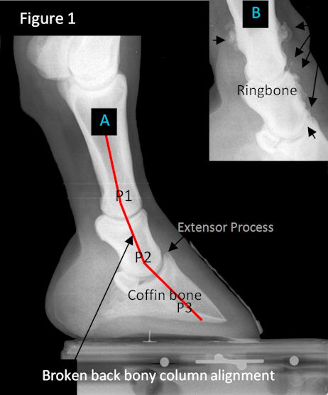
Figure 1
Note A) location of the three phalangeal bones: P1, P2, P3. The bones should be aligned in a straight line (not broken lines like they present in the image).
Without this alignment, pathology occurs such as:
- cranial joint space narrowing at the DIP and PIP joints with significant inflammation
- inflammation around the extensor process and subsequent spur formation, tendon ossification
- B) bone remodeling such as articular ringbone which forms inside the joint spacing or peri-articular ringbone which forms exterior of the joint articulation. This can cause joint fusion, and/or friction with, micro tears, and calcification, of nearby tendons/ligaments and create painful adhesions.
- thinning of sole creating demineralization of distal margin of P3
- Excess friction with navicular bursa/bone from low angles of the navicular bone due to broken back bony column alignment.
- loss of digital cushion, frog, ungual cartelige as horse avoids caudal pain with toe first landings
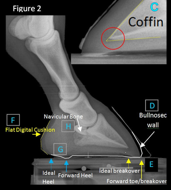
Figure 2
Observe the bottom tip of P3 (coffin bone) and notice in C)how the distal tip is resorbing (disappearing). Often the long toe creates so much leverage before the heel can lift off the ground that the horse experiences caudal heel pain and alters its gait to land toe first. Long term concussion and inflammation from a toe-first landing eventually resorbs (breaks down and reabsorbs bone) or remodels the distal tip and margins (surface) of the coffin bone.
Sometimes the hoof wall D) will look "bullnosed" where the foot is desperately trying to wear away the long toe to get rid of the leverage, but the bony column is too broken back and the foot isn't able to correct itself without intervention. Many coffin bone fractures occur from extreme stressors and hoof imbalances from long term LONG TOE UNDERRUN HEEL (broken bony column alignment) or elongated toes and forward migrated hoof capsules with excessive toe leverage and lowered palmar angles.
Additionally, the back part of the foot becomes inflamed and painful from the long toe- delayed breakover E). Circulation decreases resulting in digital cushion F) deterioration until there is little soft tissue structure left in the back part of the hoof to sustain bony column alignment. Without important support structures the heels collapse and run forward G) following the long forward toe. Eventually the navicular bone H) (which drops in angle) rubs against the deep digital flexor tendon and the friction creates inflammation which destroys the tendon sheathe, the navicular bursa, and starts to attack the navicular bone. This creates severe pain in the back part of the hoof and what is often called "Navicular Syndrome" or "Caudal Heel Syndrome".
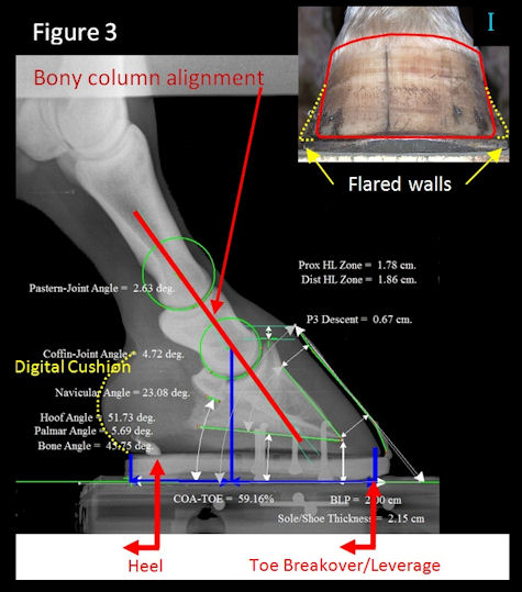
Figure 3
I) Anytime a horse has flares, this is a sign of lamellar instability (from toe leverage to inflammation of the laminae). The laminae are a bonding structure that "glues the hoof wall to the bone inside". Flares are poor hoof wall/laminae connections that are actually stretching and separating away from the coffin bone! Notice if your horse has flares how the soles flatten out. This is because as more and more flaring occurs, the hoof wall becomes unstable, and is no longer held or attached to the bone (via the laminae) so the coffin bone descends in the foot, and as it descends, the crucial healthy concave sole flattens and bottoms out. This thin sole no longer protects the sensitive corium or the circumflex artery from compressive forces under load.
The podiagry balance radiograph images shown in Figure 3 are a special technique developed by Dr. Ric Redden and have become the golden international standard for measurement of the distal limb. The use of Epona Metron software to calibrate and measure various soft and hard tissue parameters greatly aids to accomplish optimal farriery work.
Reworking the unbalanced hoof
- Trim to achieve bony column alignment of P1, P2 P3
- Trim to reduce leverage forces on hoof to achieve approx. 50/50 balance front to back from COR. (Images present 60/40
- Trim to improve (increase) navicular bone angle which reduces friction with DDFT (accomplished through bony alignment)
- Trim to improve heel base. Heel is moved back to support under the bulbs across the widest part of the frog without sacrificing heel height (no lowering of PA) otherwise add external assist as needed.
- Trim bars and clean out commissures to prevent excess pressure points and maintain heel and quarter wall integrity.
- Trim frog tags and center sulcus to prevent bacteria proliferation.
- Digital cushions are stimulated promoting healthy circulation in the back part of the foot from correct trimming and balancing of foot front to back.
- Balanced trimming promotes recovery of healthy sole concavity.


